


Showing posts with label Mesothelioma. Show all posts
Showing posts with label Mesothelioma. Show all posts
Thursday, January 30, 2014
Mesothelioma Treatment
Mesothelioma Therapy
Treatment options for mesothelioma contain surgery, chemotherapy and/or light therapy. Many mesothelioma doctors prefer to combine two or more of these treatments, an approach known as multimodal treatments. Clinical trials show this process has improved survival prices.
Surgery
Curative surgery can be obtained for early stage patients, while palliative surgery is most beneficial for late-stage patients and aids you to ease symptoms.
Chemotherapy
Chemotherapy is usually combined with surgery or maybe radiation therapy to wipe out malignant cells, shrink cancers, prevent recurrence and ease symptoms.
Radiation
Radiation therapy is used alone or in conjunction with chemotherapy or surgery to kill cancer cells, manage tumors and prevent tumor seeding.
Palliative treatments that ease symptoms are quite common for patients off stages. Experimental therapies like immunotherapy are showing progress in the future of mesothelioma treatment. Moreover, less traditional alternative treatments are offered and widely touted by mesothelioma survivors.
How Mesothelioma is Diagnosed
All patients have a unique path to a examination, but the most important factors to an accurate diagnosis are imaging runs and biopsies. Doctors use numerous tests to diagnose mesothelioma. A lot of people initially undergo a basic chest X-ray to confirm for any abnormalities. If abnormal growth is detected, a doctor will recommend an increasingly detailed imaging scan like a new PET scan, CT scan or even MRI. If mesothelioma is thought, a biopsy will be proposed. In a biopsy, a tissue sample is collected to make sure that the presence of mesothelioma cells.
Blood tests for mesothelioma may also be available, but they do not really confirm the presence of mesothelioma. Research and development is underway to discover if mesothelioma blood tests can certainly help in early diagnosis for at-risk ex - asbestos workers.

Staging
There have least five systems that doctors use with the staging of pleural mesothelioma. Older systems like those put together by Drs. Butchart and Sugarbaker didn't classify tumors with tumor-node-metastasis (TNM) descriptors, and so the International Mesothelioma Interest Group (IMIG) created the detailed IMIG staging program in 1995. This system is the preferred staging system for mesothelioma.
Blood tests for mesothelioma may also be available, but they do not really confirm the presence of mesothelioma. Research and development is underway to discover if mesothelioma blood tests can certainly help in early diagnosis for at-risk ex - asbestos workers.
Staging
There have least five systems that doctors use with the staging of pleural mesothelioma. Older systems like those put together by Drs. Butchart and Sugarbaker didn't classify tumors with tumor-node-metastasis (TNM) descriptors, and so the International Mesothelioma Interest Group (IMIG) created the detailed IMIG staging program in 1995. This system is the preferred staging system for mesothelioma.
Mesothelioma overview
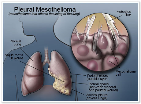
Mesothelioma is often a rare cancer caused almost exclusively by exposure to asbestos. It usually affects the thin, protective membrane surrounding the lungs, cardiovascular or abdominal cavity. Doctors diagnose approximately 3, 000 cases of mesothelioma a year in the states, and the majority of these are traced to job-related direct exposure.
Although asbestos use diminished dramatically in recent decades within this country, the incidence connected with mesothelioma remains steady. That difference can be traced to the distinct latency period linked to mesothelioma. The disease might take anywhere from 20 to be able to 50 years after exposure to asbestos before it exhibits obvious symptoms and an oncologist might make a definitive diagnosis. While no cure to the disease exists and the prognosis is commonly poor, researchers made significant progress in recent times in understanding mesothelioma and developing new therapies and alternative therapies.
Precisely how Asbestos Causes Mesothelioma
Mesothelioma cancer develops after exposure to asbestos, which most often occurs on the job – in industrial controls, shipyards, auto repair shops, old houses, schools as well as public buildings. It normally takes long-term exposure to put someone in jeopardy, asbestos is highly deadly. Even short-term and one-time exposures are recognized by cause mesothelioma cancer.
Mesothelioma Symptoms
Because early symptoms of mesothelioma usually are so mild, few people discover or recognize them, and many don't experience any kind of symptoms until later stages in the cancer. Fatigue and slight pain across the tumor may surface in first stages. Late-stage malignant mesothelioma symptoms are more noticeable and commonly provoke anyone to visit the doctor. These late-onset symptoms normally include shortness of breath, chronic pain near the tumor, weight loss, fluid build-up or bowel obstruction. Effective therapies are available to relieve symptoms, and several treatments, like talc pleurodesis, can even prevent symptom recurrence.
Thursday, January 23, 2014
diagnosis of mesothelioma
Fibrous mesothelioma diagnosis at an early stage
Early diagnosis of mesothelioma can help in effective treatment and extending the life of the patient , but as discussed , it is not easy to do so . Here are some simple tips that can really help in the early diagnosis
I know history review Alama with asbestos
If you're in the workplace current or past involved dealing with asbestos or products , you face a higher risk of asbestos exposure and mesothelioma .
If this is true , you need to take cautious of sudden weight loss , chest pain , and cough ( which lasts for a long time ) seriously.
As you can see all of them are common symptoms of mesothelioma.
If your home is near asbestos mines
If your home is located near the mine or factory asbestos , you need to be more careful and take the above symptoms seriously.
Frankly discussion with your doctor
The key point here is even more caution for common symptom if you feel you have been exposed to asbestos.
Remember, you need to consider in the past 10-20 years of history here as mesothelioma takes a lot of time to develop . Thus in the case of the occurrence of such symptoms visit your doctor , your history , said the past with asbestos and insist on checking details .
Studies and Research
British research showed that mesothelioma has many species which is pleural mesothelioma , which develops in the lining of the lungs , peritoneal mesothelioma , which develops in the lining of the abdominal cavity , pericardial mesothelioma , which affects the lining of the heart ,
Pericardial mesothelioma , which affect the testis , omental mesothelioma which is affecting the abdomen
They recognized that early diagnosis of mesothelioma can help in effective treatment and extending the life of the patient consult a doctor immediately
short note about mesothelioma
mesothelioma is a rare form of cancer is known to be caused due to exposure to asbestos minerals.
While there are several types of mesothelioma , but it is most commonly found in the lining of the lungs and abdomen .
In most patients , it was revealed mesothelioma only when it reached an advanced stage . This is because the symptoms of mesothelioma can seriously take up to 10-15 years to show up until this period, the patient did not have any symptoms related to cancer strange .
Symptoms of mesothelioma for early stage
Cold , cough , fatigue, body disorders
Types of mesothelioma
Before talking more about the symptoms of mesothelioma , it is important to know the types of mesothelioma , and some of the symptoms specific to the type of cancer.
There are five types of mesothelioma :
Pleural mesothelioma : develops in the lining of the lungs
Peritoneal mesothelioma : develops in the lining of the abdominal cavity
Pericardial mesothelioma : affects the lining of the heart
Pericardial mesothelioma : that affect testicular
Omental mesothelioma : affecting the abdomen
Symptoms of pleural mesothelioma
Pleural mesothelioma , a cancer in the lining of the lungs is the most common type of mesothelioma.
Pleural effusion
Is one of the most important symptoms of pleural mesothelioma . And pleural effusion is an accumulation of fluid between the parietal pleura and visceral pleura , as research indicates that nearly ninety percent of mesothelioma patients had pleural pleural effusion .

Shortness of breath is the second most common presentation related pleural mesothelioma
Pain in the chest / ribs during breathing
Fatigue
Dry cough
Sweating
Weight loss with no apparent reason
Symptoms of peritoneal mesothelioma
Peritoneal mesothelioma is the second most common type of mesothelioma occurs in the lining of the abdominal cavity , known as peritonitis .
As is pleural mesothelioma , where it seems that the most common symptoms and the final to be a pleural effusion , in peritoneal mesothelioma is the accumulation of fluid between the peritoneum and abdominal organs is called ascites .
While there are several types of mesothelioma , but it is most commonly found in the lining of the lungs and abdomen .
In most patients , it was revealed mesothelioma only when it reached an advanced stage . This is because the symptoms of mesothelioma can seriously take up to 10-15 years to show up until this period, the patient did not have any symptoms related to cancer strange .
Symptoms of mesothelioma for early stage
Cold , cough , fatigue, body disorders
Types of mesothelioma
Before talking more about the symptoms of mesothelioma , it is important to know the types of mesothelioma , and some of the symptoms specific to the type of cancer.
There are five types of mesothelioma :
Pleural mesothelioma : develops in the lining of the lungs
Peritoneal mesothelioma : develops in the lining of the abdominal cavity
Pericardial mesothelioma : affects the lining of the heart
Pericardial mesothelioma : that affect testicular
Omental mesothelioma : affecting the abdomen
Symptoms of pleural mesothelioma
Pleural mesothelioma , a cancer in the lining of the lungs is the most common type of mesothelioma.
Pleural effusion
Is one of the most important symptoms of pleural mesothelioma . And pleural effusion is an accumulation of fluid between the parietal pleura and visceral pleura , as research indicates that nearly ninety percent of mesothelioma patients had pleural pleural effusion .
Shortness of breath is the second most common presentation related pleural mesothelioma
Pain in the chest / ribs during breathing
Fatigue
Dry cough
Sweating
Weight loss with no apparent reason
Symptoms of peritoneal mesothelioma
Peritoneal mesothelioma is the second most common type of mesothelioma occurs in the lining of the abdominal cavity , known as peritonitis .
As is pleural mesothelioma , where it seems that the most common symptoms and the final to be a pleural effusion , in peritoneal mesothelioma is the accumulation of fluid between the peritoneum and abdominal organs is called ascites .
Wednesday, January 22, 2014
Psychosocial needs of patients of malignant Mesothelioma
Some Psychosocial needs of patients of malignant Mesothelioma
Information and communication needs
Patients diagnosed with malignant mesothelioma and their carers require clear
communication and tailored, accurate information from health professionals about
diagnosis, prognosis, treatment options and end of life issues. There is a modest amount
of evidence to suggest that adequate information is lacking in some domains.
In a UK survey of 83 patients by the British Lung Foundation, over 80% of patients
reported receiving information about treatment options, welfare benefits and
compensation; a lower proportion of patients received information about where to go for
further advice, including out of hours support (62%) and how to control symptoms (53%).
Even fewer received information about end of life issues and palliative care (25%) (301).
In a qualitative Australian study of 13 people including two patients and six carers (302),
results indicated that it was difficult to obtain reliable and accurate information relating
to the disease. When information was not provided by their health professionals, internet
searches resulted in negative or pessimistic information. Respondents also reported that
patients were referred to palliative care too late into their cancer experience (302).

It is important that patients and carers are given the right information at the right time. In
a small qualitative study of five patients with malignant mesothelioma, patients reported
not being provided with the right information and support at the right time. They were
unable to take in information due to the shock of the diagnosis and the overwhelming
amounts of information being provided (303). These findings were echoed in a UK
study of 15 patients, who recalled receiving a ‘hopeless message’ of incurable disease
with no effective treatments sympathetically delivered by doctors. Issues relating to
communication causing distress were reported to continue over the illness trajectory
Information and communication needs
Patients diagnosed with malignant mesothelioma and their carers require clear
communication and tailored, accurate information from health professionals about
diagnosis, prognosis, treatment options and end of life issues. There is a modest amount
of evidence to suggest that adequate information is lacking in some domains.
In a UK survey of 83 patients by the British Lung Foundation, over 80% of patients
reported receiving information about treatment options, welfare benefits and
compensation; a lower proportion of patients received information about where to go for
further advice, including out of hours support (62%) and how to control symptoms (53%).
Even fewer received information about end of life issues and palliative care (25%) (301).
In a qualitative Australian study of 13 people including two patients and six carers (302),
results indicated that it was difficult to obtain reliable and accurate information relating
to the disease. When information was not provided by their health professionals, internet
searches resulted in negative or pessimistic information. Respondents also reported that
patients were referred to palliative care too late into their cancer experience (302).
It is important that patients and carers are given the right information at the right time. In
a small qualitative study of five patients with malignant mesothelioma, patients reported
not being provided with the right information and support at the right time. They were
unable to take in information due to the shock of the diagnosis and the overwhelming
amounts of information being provided (303). These findings were echoed in a UK
study of 15 patients, who recalled receiving a ‘hopeless message’ of incurable disease
with no effective treatments sympathetically delivered by doctors. Issues relating to
communication causing distress were reported to continue over the illness trajectory
Difrencess benign mesothelial hyperplasia from malignant pleural mesothelioma
As emphasised earlier in this chapter, the demonstration of fat or stromal tissue invasion
by histology or imaging is an essential criterion for definitive diagnosis of malignant
pleural mesothelioma.
Although reactive mesothelial proliferations are non-invasive, entrapment of benign
mesothelial cells within the fibrous tissue of organising inflammation can simulate
neoplastic invasion . This can make histological discrimination between
entrapment and invasion difficult. It is recommended that when invasion cannot be
identified in biopsy tissue, the lesion should be designated as an atypical mesothelial
proliferation
Clinical decision-making for a diagnosis of malignant mesothelioma may be made when
a limited biopsy has shown an atypical mesothelial proliferation without invasion. This
requires correlation with imaging studies, a more adequate biopsy or, in many instances,
serial imaging studies to ascertain whether the lesion is progressive
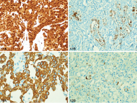
Immunohistochemistry in the diagnosis of malignant mesothelioma
Immunohistochemistry is integral to the diagnosis of malignant mesothelioma and
is currently the most useful and standard ancillary procedure for distinguishing this
malignancy from other types of cancer.
The primary differential diagnosis for epithelioid mesothelioma in the pleura is with
metastatic lung adenocarcinoma. Immunohistochemistry has replaced electron
microscopy as the preferred ancillary method, and differential diagnosis now relies on
the detection of various mesothelial and carcinoma-related antigens/markers in cytology
cell block sections or in biopsy tissue (21, 40, 41, 44, 45, 63, 78, 79). Carcinoma-related
markers include carcinoembryonic antigen (CEA), LeuM1 (CD15), Ber-EP4, B72.3 and
BG8 (45, 63, 80-84) and – whenever lung adenocarcinoma is included in the differential
diagnosis – thyroid transcription factor-1 (TTF-1) (45) and/or napsin A (85, 86). Antigens
characteristically expressed by mesothelial cells include calretinin, Wilms’ tumour gene
product (WT-1), mesothelin, cytokeratin 5/6, HBME-1 antigen, thrombomodulin and
podoplanin (D2-40) antibody (63, 79, 87-113).
The exact combination and number of antigens to evaluate is dependent on the
differential diagnosis and the antibodies available. Currently, calretinin is considered
to have the greatest specificity for a diagnosis of malignant mesothelioma, followed
by WT1 and D2-40 (21, 44, 45, 79, 99). The International Mesothelioma Panel (IMP) (41)
recommends at least one cytokeratin (CK) marker plus at least two mesothelial markers
(for example, calretinin and WT1) together with at least two carcinoma-related markers
(for example, CD15 and TTF-1). The guidelines from the ERS and the ESTS (21) reiterate
this IMP approach, as do the Guidelines from the International Mesothelioma Interest
Group (IMIG)(45). When tumours other than lung cancer enter into the differential
diagnosis (for example, secondary prostate carcinoma) additional markers become
necessary. The ERS/ESTS guidelines do not recommend use of CK7/CK20 (114) for
diagnosis of mesothelioma (21).
As a practical reference for pathologists, the IMIG recommends that markers have
sensitivity or specificity greater than 80% for the lesions in question (45), whereas the
ERS/ESTS guidelines specify a minimum sensitivity of 60-70%. Interpretation of positivity
should take into account the localisation of the stain (for example, nuclear versus
cytoplasmic) and the percentage of cells stained: more than 10% has been suggested for
cytoplasmic membranous markers (45).
From the preceding discussion, it is clear that none of the antibodies used for the
diagnosis of mesothelioma is 100% specific or sensitive – hence the requirement for
panels of mesothelial and non-mesothelial antibodies. As one example of the diagnostic
pitfalls that can be encountered, up to 15% of a subset of high-grade carcinomas of the
breast can express calretinin, and these carcinomas may also express CK5/6 and lack
detectable oestrogen receptor protein – with the potential for misdiagnosis of pleural
metastases as malignant mesothelioma (115, 116).
Immunohistochemistry has a more restricted role for the diagnosis of sarcomatoid
malignant mesotheliomas than for malignant mesotheliomas with an epithelial
30
component, because many sarcomatoid malignant mesotheliomas express only
cytokeratins in addition to vimentin and, in some cases, smooth muscle markers
Expression of calretinin is variable (30-89%) in sarcomatoid areas of
mesothelioma. . The high percentage labelling recorded in some
studies is explicable by acceptance of cytoplasmic labelling for calretinin as a positive
result (117), whereas positive nuclear labelling is required in addition to any cytoplasmic
labelling (41, 44). Most sarcomatoid and desmoplastic malignant mesotheliomas
are strongly positive for cytokeratins (although CK-negative sarcomatoid malignant
mesotheliomas do occur), and CK labelling can also highlight invasion, such as genuine
invasion into subpleural fat by a desmoplastic malignant mesothelioma . The ERS/
ESTS guidelines recommend use of at least two broad-spectrum CK antibodies and
two markers with negative predictive value, to support a diagnosis of sarcomatoid
mesothelioma

The place of immunohistochemistry in the diagnosis of malignant pleural mesothelioma
is a constantly evolving area and specific information on antibodies and their source
should be obtained from the current literature. It also seems likely that molecular
approaches to diagnosis – such as profiling of microRNA expression in tumour
tissue or extrapleural samples – will supplement immunohistochemistry for the
diagnosis of mesothelioma, but these approaches are at an investigational phase of
evaluation and at present they cannot be recommended for routine use in diagnosis.
is currently the most useful and standard ancillary procedure for distinguishing this
malignancy from other types of cancer.
The primary differential diagnosis for epithelioid mesothelioma in the pleura is with
metastatic lung adenocarcinoma. Immunohistochemistry has replaced electron
microscopy as the preferred ancillary method, and differential diagnosis now relies on
the detection of various mesothelial and carcinoma-related antigens/markers in cytology
cell block sections or in biopsy tissue (21, 40, 41, 44, 45, 63, 78, 79). Carcinoma-related
markers include carcinoembryonic antigen (CEA), LeuM1 (CD15), Ber-EP4, B72.3 and
BG8 (45, 63, 80-84) and – whenever lung adenocarcinoma is included in the differential
diagnosis – thyroid transcription factor-1 (TTF-1) (45) and/or napsin A (85, 86). Antigens
characteristically expressed by mesothelial cells include calretinin, Wilms’ tumour gene
product (WT-1), mesothelin, cytokeratin 5/6, HBME-1 antigen, thrombomodulin and
podoplanin (D2-40) antibody (63, 79, 87-113).
The exact combination and number of antigens to evaluate is dependent on the
differential diagnosis and the antibodies available. Currently, calretinin is considered
to have the greatest specificity for a diagnosis of malignant mesothelioma, followed
by WT1 and D2-40 (21, 44, 45, 79, 99). The International Mesothelioma Panel (IMP) (41)
recommends at least one cytokeratin (CK) marker plus at least two mesothelial markers
(for example, calretinin and WT1) together with at least two carcinoma-related markers
(for example, CD15 and TTF-1). The guidelines from the ERS and the ESTS (21) reiterate
this IMP approach, as do the Guidelines from the International Mesothelioma Interest
Group (IMIG)(45). When tumours other than lung cancer enter into the differential
diagnosis (for example, secondary prostate carcinoma) additional markers become
necessary. The ERS/ESTS guidelines do not recommend use of CK7/CK20 (114) for
diagnosis of mesothelioma (21).
As a practical reference for pathologists, the IMIG recommends that markers have
sensitivity or specificity greater than 80% for the lesions in question (45), whereas the
ERS/ESTS guidelines specify a minimum sensitivity of 60-70%. Interpretation of positivity
should take into account the localisation of the stain (for example, nuclear versus
cytoplasmic) and the percentage of cells stained: more than 10% has been suggested for
cytoplasmic membranous markers (45).
From the preceding discussion, it is clear that none of the antibodies used for the
diagnosis of mesothelioma is 100% specific or sensitive – hence the requirement for
panels of mesothelial and non-mesothelial antibodies. As one example of the diagnostic
pitfalls that can be encountered, up to 15% of a subset of high-grade carcinomas of the
breast can express calretinin, and these carcinomas may also express CK5/6 and lack
detectable oestrogen receptor protein – with the potential for misdiagnosis of pleural
metastases as malignant mesothelioma (115, 116).
Immunohistochemistry has a more restricted role for the diagnosis of sarcomatoid
malignant mesotheliomas than for malignant mesotheliomas with an epithelial
30
component, because many sarcomatoid malignant mesotheliomas express only
cytokeratins in addition to vimentin and, in some cases, smooth muscle markers
Expression of calretinin is variable (30-89%) in sarcomatoid areas of
mesothelioma. . The high percentage labelling recorded in some
studies is explicable by acceptance of cytoplasmic labelling for calretinin as a positive
result (117), whereas positive nuclear labelling is required in addition to any cytoplasmic
labelling (41, 44). Most sarcomatoid and desmoplastic malignant mesotheliomas
are strongly positive for cytokeratins (although CK-negative sarcomatoid malignant
mesotheliomas do occur), and CK labelling can also highlight invasion, such as genuine
invasion into subpleural fat by a desmoplastic malignant mesothelioma . The ERS/
ESTS guidelines recommend use of at least two broad-spectrum CK antibodies and
two markers with negative predictive value, to support a diagnosis of sarcomatoid
mesothelioma
The place of immunohistochemistry in the diagnosis of malignant pleural mesothelioma
is a constantly evolving area and specific information on antibodies and their source
should be obtained from the current literature. It also seems likely that molecular
approaches to diagnosis – such as profiling of microRNA expression in tumour
tissue or extrapleural samples – will supplement immunohistochemistry for the
diagnosis of mesothelioma, but these approaches are at an investigational phase of
evaluation and at present they cannot be recommended for routine use in diagnosis.
Cytological features of malignant mesothelioma
The majority opinion among surgical pathologists is that an essential condition for
definitive histological diagnosis of pleural mesothelioma is the demonstration of
neoplastic invasion – such as infiltration into underlying fat, skeletal muscle, rib or lung –
as opposed to benign entrapment of mesothelium
Effusion fluid cytology in isolation does not allow assessment of invasion, although a
2007 Update Statement on Mesothelioma from the British Thoracic Society (BTS)
stated that cytological examination of pleural effusion fluid from patients may be
sufficient for diagnosis in some patients, when correlated with imaging studies – that is,
using imaging studies as a surrogate for the histological demonstration of invasion
For example, the combination of the following may allow a diagnosis of mesothelioma
at a high level of confidence: florid atypical mesothelial proliferation on pleural effusion
fluid cytology supported by immunohistochemical studies on cell-block sections and
with no evidence of any infective process on microbiological investigation, plus confluent
pleural thickening with nodularity on imaging studies (with/without evidence of chest
wall invasion), plus absence from imaging studies of any intrapulmonary mass lesion or
extrathoracic tumour with the capacity for spread to the pleura.

Cytology-only diagnosis based on effusion fluids remains controversial Although
several cytological findings raise varying levels of suspicion of malignant pleural
mesothelioma (58) – such as the extent of the mesothelial proliferation, the presence of
papillary structures (especially in the pleura), cytological atypia, frequent cytoplasmic
vacuoles and focal necrosis – there is some overlap in the cytological appearances
between reactive mesothelial hyperplasia and malignant mesothelioma
The most useful cytological features of malignant mesothelioma include the presence
of numerous relatively large (>50 cell) balls of cells with berry-like external contours
comprising cells that are much larger (with enlarged cytoplasm, nucleus and nucleolus)
than most benign mesothelial cells; the presence of macronucleoli – although prominent
nucleoli can be present in reactive mesothelial cells and not all malignant mesothelioma
cells have macronucleoli; and nuclear atypia.
Many cytological features of malignant mesothelioma – such as scalloped borders of cell
clumps, intercellular windows, variation in cytoplasmic staining and its ‘density’, and low
nuclear-to-cytoplasmic ratios – are shared between reactive and malignant epithelioid
mesothelial cells .
Reported sensitivities for a clear cytodiagnosis of mesothelioma on effusion fluids have
ranged widely. One 1997 study reported a low sensitivity of 32% ). In another study
26
of 162 cases , effusion fluid cytology showed high specificity (~99%) when all criteria
specified for mesothelioma were fulfilled, but the sensitivity was only 47.5% when
not all criteria were met. This sensitivity was improved by interpreting the cytological
findings together with effusion fluid hyaluronic acid concentrations. Some centres with
specialised interest and experience in the cytodiagnosis of mesothelioma from effusion
fluid (58) have found a high positive predictive value for diagnosis. Such results may
not be obtainable for other centres with less experience in cytological assessment of
mesothelial proliferations.
definitive histological diagnosis of pleural mesothelioma is the demonstration of
neoplastic invasion – such as infiltration into underlying fat, skeletal muscle, rib or lung –
as opposed to benign entrapment of mesothelium
Effusion fluid cytology in isolation does not allow assessment of invasion, although a
2007 Update Statement on Mesothelioma from the British Thoracic Society (BTS)
stated that cytological examination of pleural effusion fluid from patients may be
sufficient for diagnosis in some patients, when correlated with imaging studies – that is,
using imaging studies as a surrogate for the histological demonstration of invasion
For example, the combination of the following may allow a diagnosis of mesothelioma
at a high level of confidence: florid atypical mesothelial proliferation on pleural effusion
fluid cytology supported by immunohistochemical studies on cell-block sections and
with no evidence of any infective process on microbiological investigation, plus confluent
pleural thickening with nodularity on imaging studies (with/without evidence of chest
wall invasion), plus absence from imaging studies of any intrapulmonary mass lesion or
extrathoracic tumour with the capacity for spread to the pleura.
Cytology-only diagnosis based on effusion fluids remains controversial Although
several cytological findings raise varying levels of suspicion of malignant pleural
mesothelioma (58) – such as the extent of the mesothelial proliferation, the presence of
papillary structures (especially in the pleura), cytological atypia, frequent cytoplasmic
vacuoles and focal necrosis – there is some overlap in the cytological appearances
between reactive mesothelial hyperplasia and malignant mesothelioma
The most useful cytological features of malignant mesothelioma include the presence
of numerous relatively large (>50 cell) balls of cells with berry-like external contours
comprising cells that are much larger (with enlarged cytoplasm, nucleus and nucleolus)
than most benign mesothelial cells; the presence of macronucleoli – although prominent
nucleoli can be present in reactive mesothelial cells and not all malignant mesothelioma
cells have macronucleoli; and nuclear atypia.
Many cytological features of malignant mesothelioma – such as scalloped borders of cell
clumps, intercellular windows, variation in cytoplasmic staining and its ‘density’, and low
nuclear-to-cytoplasmic ratios – are shared between reactive and malignant epithelioid
mesothelial cells .
Reported sensitivities for a clear cytodiagnosis of mesothelioma on effusion fluids have
ranged widely. One 1997 study reported a low sensitivity of 32% ). In another study
26
of 162 cases , effusion fluid cytology showed high specificity (~99%) when all criteria
specified for mesothelioma were fulfilled, but the sensitivity was only 47.5% when
not all criteria were met. This sensitivity was improved by interpreting the cytological
findings together with effusion fluid hyaluronic acid concentrations. Some centres with
specialised interest and experience in the cytodiagnosis of mesothelioma from effusion
fluid (58) have found a high positive predictive value for diagnosis. Such results may
not be obtainable for other centres with less experience in cytological assessment of
mesothelial proliferations.
diagnosis of malignant mesothelioma
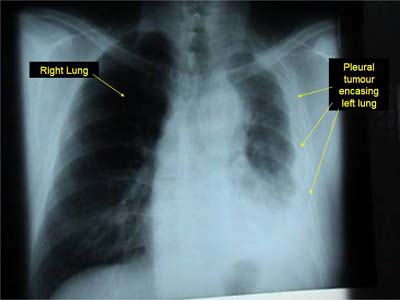
The diagnosis of malignant mesothelioma can be difficult, with symptoms and clinical
findings that can mimic and be mimicked by other diseases. Pleural mesothelioma
patients may present with dyspnoea, chest pain (pleuritic or non-pleuritic), cough
and weight loss, or any combinations of these symptoms (39-42). Initial clinical and
radiological examination usually reveals a pleural effusion, often massive. Rarely,
patients are asymptomatic at the time when a radiological abnormality is demonstrated,
and patients seldom present with metastatic disease.
Some patients with malignant mesothelioma experience a long interval between the
first onset of symptoms and subsequent diagnosis, but whether a long interval signifies
enhanced or diminished survival following diagnosis is unclear. Most patients with
malignant pleural mesothelioma have a background of asbestos exposure (40, 42), and
some may have had antecedent symptoms associated with benign asbestos-related
disease – for example, symptoms related to asbestosis or benign asbestos pleuritis with
effusion. Others may have radiological evidence of past asbestos exposure, such as
pleural plaques.
In general, biopsy, immunohistochemical analysis and correlation with radiological
and clinical features are needed for the diagnosis of mesothelioma (42). When
immunohistochemical findings are non-diagnostic or discordant, electron microscopy
– including electron microscopic examination of tissue retrieved from blocks of paraffinembedded
biopsy tissue or cytology cell blocks – can be used, but electron microscopy is
not recommended for ‘routine’ diagnosis of mesothelioma (21, 43).
Although several cytological and histological findings may raise varying levels of
suspicion of malignant pleural mesothelioma (see section 2.4) a current requirement
for the definitive clinicopathological diagnosis of malignant pleural mesothelioma is the
demonstration of neoplastic invasion – for example, infiltration into subpleural fat, chest
wall skeletal muscle, rib or lung – by histological examination or by imaging studies,
and by clinical exclusion of alternative causes for an atypical mesothelial
proliferation.
A component of malignant mesothelioma in situ can be diagnosed when invasion has
been demonstrated in the same or different biopsy or by imaging studies (44). This
applies specifically to epithelioid malignant mesotheliomas. Sarcomatoid malignant
mesotheliomas are rarely diagnosable from effusion fluid cytology and are usually
identified histologically, by the demonstration of invasion or overtly sarcomatoid areas.
Incidence of malignant mesothelioma
Variation in the incidence of malignant mesothelioma is reported in different parts
of the world. For example, seven people per million in Japan have been diagnosed
with malignant mesothelioma compared with 40 people per million in Australia.
These differences are largely attributable to the amount of asbestos ‘consumed’ in
a certain period (25).
Australia, as one of the largest consumers of asbestos worldwide in the post-World War
II period, has one of the highest incidences of malignant mesothelioma. Around 660 new
cases of malignant mesothelioma were documented in 2007 and, in terms of mortality,
this disease is approaching the numbers of deaths caused by multiple myeloma and
ovarian cancer.
There is also regional variation in the incidence of malignant mesothelioma. For example,
in Australia the highest reported incidence has been in men in Western Australia. This
variation is largely attributable to occupational exposure associated with crocidolite
mining in Wittenoom
Most deaths caused by malignant mesothelioma in Australia and other developed
countries are due to occupational exposure to asbestos. The frequency of cases
attributable to occupational exposure may have begun to decline owing to stringent
control of asbestos use and handling. Asbestos, however, persists in our natural and
built environments, and it is important that we continue to minimise exposure to it
by all reasonable means. Among mesothelioma patients who do not have a history of
occupational exposure, there is now a high proportion of people with a history of home
renovation, in which exposure to asbestos might have occurred (26). Research is needed
to determine if asbestos exposure explains this high proportion. It is important also that
we remain alert to sources of possible exposure to asbestos in the community and control
any such exposure as it is identified.
Data on the incidence and mortality of malignant mesothelioma in Aboriginal and Torres
Strait Islanders and culturally and linguistically diverse groups has not been reliably
estimated due to the lack of recorded ethnicity. However, from July 2010, all new cases
of malignant mesothelioma diagnosed in Australia are monitored by the Australian
Mesothelioma Registry.
OVERVIEW OF A MESOTHELIOMA LAWSUIT
Because of the aggressive nature of Mesothelioma cancer, a
victim has limited time to spend with loved ones. A diagnosis of
any asbestos-related disease is financially draining as well as
emotionally exhausting. Consider that a Mesothelioma lawsuit is
an important way to fund costly treatment options for victims and
to provide vital financial security down the road.
The first step after receiving such a life-altering diagnosis is to
seek expert medical care and a strong emotional support
network to aid in the uphill fight against the malignancy. Quality
healthcare is important in managing both the physical symptoms
of the disease and the emotional grief that accompanies a terminal
illness.
The second step should be to contact a qualified Mesothelioma
attorney who is experienced in the complexities of asbestosrelated
law and who can help make the negligent industry take
responsibility for its greed. A lawyer specializing in Mesothelioma
cases will know how to establish and prove a victim’s exposure
history, which is often essential information for a successful
lawsuit. Don’t let time run out before you fight for what you
deserve.
mesothelioma lawyers guide
INTRODUCTION
Mesothelioma cancer is a devastating but mostly preventable disease. You and your family have the right to seek compensation for this
preventable disease. Mega million dollar corporations have set aside huge sums of money to provide to victims and/or their families for
asbestos exposure related diseases.
If you are a grieving family member or executor of the will of a person who has died from asbestos-related disease or Mesothelioma,
you may be eligible to file a claim as well. Anapol Schwartz Mesothelioma lawyers can help you.
The complex maze of legal details is most likely the last thing that you want to confront after learning about an asbestos-related illness.
Yet, it’s to your advantage to let the law firm of your choice to fight for what’s right in order to create the necessary funds to finance
aggressive treatment, pay for huge medical bills incurred during diagnosis, and provide financial security for your family’s future.
The burden of proof lies on your shoulders. It is a difficult and time-consuming responsibility which is why the guidance of an experienced
Mesothelioma lawyer is critical in helping victims and your families receive financial compensation.
A good Mesothelioma attorney understands the legal complexities involved in this kind of lawsuit, including asbestos product identification,
specific asbestos-related medical issues, and specific time constraints that narrow the window of opportunity in order to file a claim.
Now is the time to find the right Mesothelioma lawyer before your state’s statutes of limitations expires, which would unfortunately leave
you and your family grieving and empty-handed.
you can download the guide from here
mesothelioma in briefs
What is Mesothelioma?
Mesothelioma is a rare type of cancer caused by asbestos exposure that affects the lining of the lungs, abdomen, and heart.
What are the symptoms of Mesothelioma?
Mesothelioma symptoms include shortness of breath, chest pain, or coughing up blood. Mesothelioma is difficult to diagnose
because it shares symptoms with many different conditions and/or natural signs of aging. Also, different types of Mesothelioma
cause different symptoms.
What should people do if diagnosed with an asbestos-related disease?
Victims should follow all their doctors’ orders. Victims should also contact an attorney to see if they have reason to pursue legal
action against the corporation responsible for their injuries.
Are there time constraints for filing a Mesothelioma suit?
Yes, individual states have laws called statues of limitations, which limit how
much time victims can initiate legal action. Do not delay.
What does the compensation cover?
The potential compensation can cover mounting medical bills resulting from tests
and treatment for your illness, pain and suffering you have experienced as well
as the mental anguish and grief suffered by you and your family, and financial
security for your family after you pass away
Monday, January 20, 2014
Clinical Picture of mesothelioma
Signs and symptoms of mesothelioma
Early symptoms of mesothelioma are more often caused by other things, so at first people
may ignore them or mistake them for common, minor ailments. Most people with
mesothelioma have symptoms for at least a few months before they are diagnosed.
Symptoms of pleural mesothelioma (mesothelioma of the chest) can include:
· Pain in the lower back or at the side of the chest
· Shortness of breath
· Fluid in the area around the lung
· Cough
· Fever
· Excessive sweating
· Fatigue
· Weight loss (without trying)
· Trouble swallowing (feeling like food gets stuck)
· Hoarseness
· Swelling of the face and arms
Symptoms of peritoneal mesothelioma can include:
· Abdominal (belly) pain
· Swelling or fluid in the abdomen
· Weight loss (without trying)
· Nausea and vomiting
The symptoms and signs above may be caused by mesothelioma, but more often they are
caused by other conditions. Still, if you have any of these problems (especially if you
have been exposed to asbestos), it's important to see your doctor right away so the cause
can be found and treated, if needed.
Sunday, January 19, 2014
mesothelial Benign tumors
Benign tumors of the mesothelium
Benign (non-cancerous) tumors can also start in the mesothelium. These tumors are
typically removed by surgery, and there is often no need for additional treatment.
Localized fibrous tumor of the pleura
This type of benign tumor can form in the pleura surrounding the lungs. It used to be
called benign fibrous mesothelioma, but doctors now know that this tumor actually starts
from tissue under the mesothelium and not from mesothelial cells. This disease is usually
benign, but about 1 in 10 are cancerous. A similar condition that starts in the peritoneum
is called solitary fibrous tumor of the peritoneum.
Adenomatoid mesothelioma
This benign tumor can develop in the mesothelium of certain reproductive organs. In
men, it often starts in the epididymis (ducts that carry sperm cells out of the testicle). In
women, this tumor may begin in the fallopian tubes (tubes that carry eggs from the
ovaries to the uterus).
Benign cystic mesothelioma
This rare non-cancerous tumor often begins in the peritoneum.
Malignant mesothelioma
Malignant mesothelioma
A cancerous tumor of the mesothelium is called a malignant
mesothelioma, although this
is often shortened to just mesothelioma. Mesotheliomas can start in 4 main areas in the
body.
· Pleural mesotheliomas start in the chest. About 3 out of 4 mesotheliomas are pleural
mesotheliomas.
· Peritoneal mesotheliomas begin in the abdomen. They make up most of the
remaining cases.
· Pericardial mesotheliomas start in the covering around the heart and are very rare.
· Mesotheliomas of the tunica vaginalis are very rare tumors that start in the covering
layer of the testicles.
Malignant mesotheliomas can also be classified into 3 main types based on how the cells
are arranged:
· About 50% to 60% of mesotheliomas are epithelioid. This type tends to have a better
outlook (prognosis) than the other types.
· About 10% to 20% of mesotheliomas are sarcomatoid (fibrous).
· Mixed (biphasic) mesotheliomas have both epithelioid and sarcomatoid areas. They
make up about 30% to 40% of mesotheliomas.
What is malignant mesothelioma
Introduction to cancer
What is cancer?
The body is made up of trillions of living cells. Normal body cells grow, divide into new
cells, and die in an orderly way. During the early years of a person's life, normal cells
divide faster so the person can grow. After the person becomes an adult, most cells divide
only to replace worn-out or dying cells or to repair injuries.
Cancer begins when cells in a part of the body start to grow out of control. There are
many kinds of cancer, but they all start because of out-of-control growth of abnormal
cells.
Cancer cell growth is different from normal cell growth. Instead of dying, cancer cells
continue to grow and form new, abnormal cells. Cancer cells can also invade (grow into)
other tissues, something normal cells cannot do. Growing out of control and invading
other tissues is what makes a cell a cancer cell.
Cells become cancer cells because of damage to DNA. DNA is in every cell and directs
all its actions. In a normal cell, when DNA gets damaged the cell either repairs the
damage or the cell dies. In cancer cells, the damaged DNA is not repaired, but the cell
doesn't die like it should. Instead, this cell goes on making new cells that the body does
not need. These new cells will all have the same damaged DNA as the first cell does.
People can inherit damaged DNA, but most DNA damage is caused by mistakes that
happen while the normal cell is reproducing or by something in our environment.
Sometimes the cause of the DNA damage is something obvious, like cigarette smoking.
But often no clear cause is found.
In most cases the cancer cells form a tumor. Some cancers, like leukemia, rarely form
tumors. Instead, these cancer cells involve the blood and blood-forming organs and
circulate through other tissues where they grow.
Cancer cells often travel to other parts of the body, where they begin to grow and form
new tumors that replace normal tissue. This process is called metastasis. It happens when
the cancer cells get into the bloodstream or lymph vessels of our body.
No matter where a cancer spreads, it is always named for the place where it started. For
example, breast cancer that has spread to the liver is still called breast cancer, not liver
cancer. Likewise, prostate cancer that has spread to the bone is metastatic prostate cancer,
not bone cancer.
Different types of cancer can behave very differently. For example, lung cancer and
breast cancer are very different diseases. They grow at different rates and respond to
different treatments. That is why people with cancer need treatment that is aimed at their
particular kind of cancer.
Not all tumors are cancerous. Tumors that aren't cancer are called benign. Benign tumors
can cause problems -- they can grow very large and press on healthy organs and tissues.
But they cannot grow into (invade) other tissues. Because they can't invade, they also
can't spread to other parts of the body (metastasize). These tumors are almost never life
threatening
.
What is malignant mesothelioma?
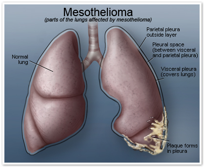
Malignant mesothelioma is a cancer that starts in cells in the linings of certain parts of the
body, especially the chest cavity or abdominal cavity..
A layer of specialized cells called mesothelial cells lines the inside of the chest, the
abdomen, and the space around your heart. These cells also cover the outer surface of
most of your internal organs. The lining formed by these cells is called the mesothelium.
The mesothelium helps protect your organs by making a special lubricating fluid that
allows organs to move. For example, this fluid makes it easier for your lungs to move
(expand and contract) inside the chest when you breathe. The mesothelium has different
names in different parts of the body:
· The pleura coats the lungs and the cavity containing the lungs in the chest.
· The peritoneum coats the abdominal cavity and many of the organs within that cavity.
· The tunica vaginalis coats the testicles.
· The pericardium coats the heart and creates the cavity that holds the heart in the
chest.
Mesothelial tumors can be non-cancerous (benign) or cancerous (malignant).
Introduction & History of Mesothelioma
Introduction
Background
Malignant mesothelioma is an aggressive tumor that originates in the serosal
membranes that line the thoracic and abdominal cavities. This disease has become
an important health issue over recent years, with Australia having one of the highest
reported incidences (2-4). More than 90% of reported cases of mesothelioma occur in
the pleura, compared with 4–7% affecting the peritoneum, and fewer than 1% jointly
occurring in the pericardium and tunica vaginalis testis (2, 4, 5). Even rarer cases have
been recorded as apparently primary ovarian mesotheliomas ).
The occurrence of malignant mesothelioma is typically related to exposure to mineral
fibres such as asbestos and erionite (8-10). Asbestos is a collection of naturally occurring
crystalline hydrated silicates that are resistant to high temperatures and humidity.
Asbestos fibres are biopersistent (retained in the human body) and can be detected as
asbestos bodies’ in the lung many years after inhalation ).
The World Health Organization (WHO) has recognised asbestos as one of the most
important occupational carcinogens and in 2010 upgraded its global estimate of asbestosrelated
diseases to 107,000 annual deaths ).
History of mesothelioma
The first studies on the association between asbestos and malignant mesothelioma
appeared in the 1950s. Weiss’ case report of asbestosis and pleural malignancy and Van
der Schoot’s paper describing three insulation workers with malignant disease were the
first of many to be published (13, 14). Wagner confirmed the association between asbestos
and malignant mesothelioma through his work in the 1950s in South Africa,
Because most asbestos exposure occurred in the work environment, malignant
mesothelioma has traditionally been considered an occupational disease. Paraoccupational
malignant mesothelioma has been described in households of asbestos
workers in which cohabitants had been exposed via contaminated clothes (16). The term
‘environmental malignant mesothelioma’ has been used to describe disease identified in
people living close to asbestos mines or factories or when people have been exposed to
asbestos or asbestos-like material present in the soil (17, 18).
Other factors have been recognised as potential causes of malignant mesothelioma.
Radiotherapy to the chest has been reported but the number of patients with this
association is limited (19). The role of SV40 (one of the simian viruses) viral infection as an
important etiologic cofactor in malignant mesothelioma remains under discussion (20, 21).
Exposure to asbestos is more common in occupations with a predominantly male workforce,
which explains why the current incidence of malignant mesothelioma is higher among men
than women. Most mesothelioma patients have been primary asbestos workers or people
who handled raw asbestos in the mining, milling, transportation and manufacturing of the
material. As this high-risk occupational exposure has been limited by the total ban on the
use of asbestos products in Australia, the exposure-mix may change to include a greater
proportion of people who have been exposed in non-occupational settings.
13
| Guidelines for the Diagnosis and Treatment of Malignant Pleural Mesothelioma |
A dose-response relationship between cumulative asbestos exposure (increased levels or
duration of exposure, or both) and malignant mesothelioma has been demonstrated ).
A ‘safe’ threshold of cumulative exposure, below which there is no increased risk, has not
been defined.
The latency period, or the period between first exposure to asbestos and the diagnosis
of mesothelioma, shows a wide range (20–60 years) and there are indications that the
latency in Australia has increased in recent years . The median age at diagnosis of
malignant mesothelioma in Australia is slightly above 70 years, with many patients
presenting with co-morbidities


